• Introduction
  • Service items
  • Our facilities
  • Health Education Content(Download)
  • Nuclear medicine is a safe and noninvasive (besides usual IV injection) imaging technology, which uses very small amounts of specially formulated radioactive materials (commonly called “tracers”), typically injected into the bloodstream or other routes by inhaling or swallowing to be intaken into the body for a time interval enough to acquire images for diagnosis. The radiotracer travels through the areas being examined and gives off energy in gamma-rays detected by a special camera, followed by computerization to create images of the related part.
  • See the scheme below:

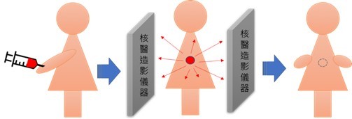
    Figure 1: The radioactive tracer is first injected into the body, and the cameras detect the released energy (gamma-rays or others), create pictures offering details on both the structure and function of the organs and tissues in the body. The small amount of radiotracer in the body will lose its radioactivity over time through the natural process of radioactive decay. (Rf. AEC.gov.tw; Atomic Energy Council, Executive Yuan, Taiwan)
  • Commonly used nuclear medicine applications include:
  1. Heart scans
  2. Bone scans
  3. Renal scans
  4. Thyroid scans
  5. Brain scans
  • Nuclear medicine is particularly valuable in providing physicians with information on both the structure and function of a patient's cells, tissue, or organs. The use of nuclear medicine procedures has kept this imaging technology in high demand. Our hospital has specially set up the Nuclear Medicine Department to help assess and diagnose disease conditions.
  • Stress & redistribution myocardial perfusion scan with SPECT, AKA MPI)

    Myocardial perfusion images (MPI) are used to evaluate coronary artery blood flow, showing how well the blood flows through the heart muscle. Furthermore, the technique can evaluate the prognosis of coronary artery surgery.

  • Whole body bone scan

    Bone scan can help diagnose a number of bone conditions, including bone cancer or skeletal metastasis, localization of bone/joint inflammation and fractures, bone infection, and others.

  • Sialoscintigraphy

    Salivary gland scintigraphy, also called sialoscintigraphy, can help diagnose and assess some salivary excretion function or disorders, including Sjögren’s syndrome (SICCA syndrome) and dry mouth in association with other autoimmune diseases.

  • Brain scan using (Tc-99m Trodat-1) for Parkinson Disease (PD)

    By injecting this tracer into the human body, the brain scan can help evaluate a brain chemical-receptor interaction involving the development movement control problems such as Parkinson's disease.

Scheduling Request: (02)2672-3456 ext. 6006
Please feel free to contact us if you need any further information.

  • SPECT

    We offer single-photon emission computed tomography (SPECT) service. The facility is a new model last updated in 2015 and first used in Taiwan in 2016.

    The images are acquired from multiple angles, usually encompassing 180 or 360 degrees around the body. These multiple-angle data sets are used to produce computer reconstructed tomographic images that can be displayed as coronal, sagittal, and transverse slices.

    Imaging Principles of SPECT:
    Projection: Acquire multiple 2-D images from multiple angles. A reconstruction algorithm obtains the 3-D images. Computer is then used to demonstrate the reconstructed slices by multiple projections.

©2026行天宮醫療志業醫療財團法人恩主公醫院 En Chu Kong Hospital. All rights reserved. Web Design by FT
Top